Your heart and how it works
The heart is the organ that pumps blood through the lungs and to the rest of the body. It is located in the chest cavity behind the breastbone, also known as the sternum.
The chambers of the heart
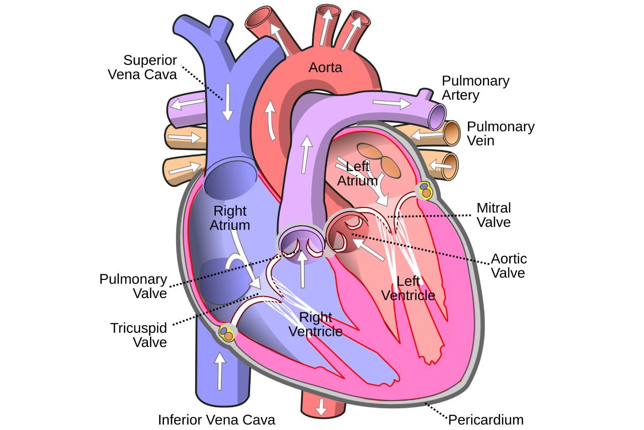
The heart is the size of your fist and is divided into four chambers named the:
- Right atrium
- Right ventricle
- Left atrium
- Left ventricle
Blood circulates throughout your body, bringing oxygen to your cells and removing carbon dioxide. Once the oxygen has been used, the blood comes back to the heart. The right atrium receives blood from the body without oxygen (unoxygenated blood). The blood in the right atrium is pumped into the right ventricle, which then pumps the blood to the lungs.
Carbon dioxide (a waste product in the blood) is removed in the lungs, and new oxygen is added. The blood then flows into the left atrium, which pumps the blood with oxygen (oxygenated blood) into the left ventricle. The left ventricle pumps blood through a blood vessel called the aorta, and out to the rest of your body.
OpenStax College, CC BY 3.0 (https://creativecommons.org/licenses/by/3.0), via Wikimedia Commons
The valves of the heart
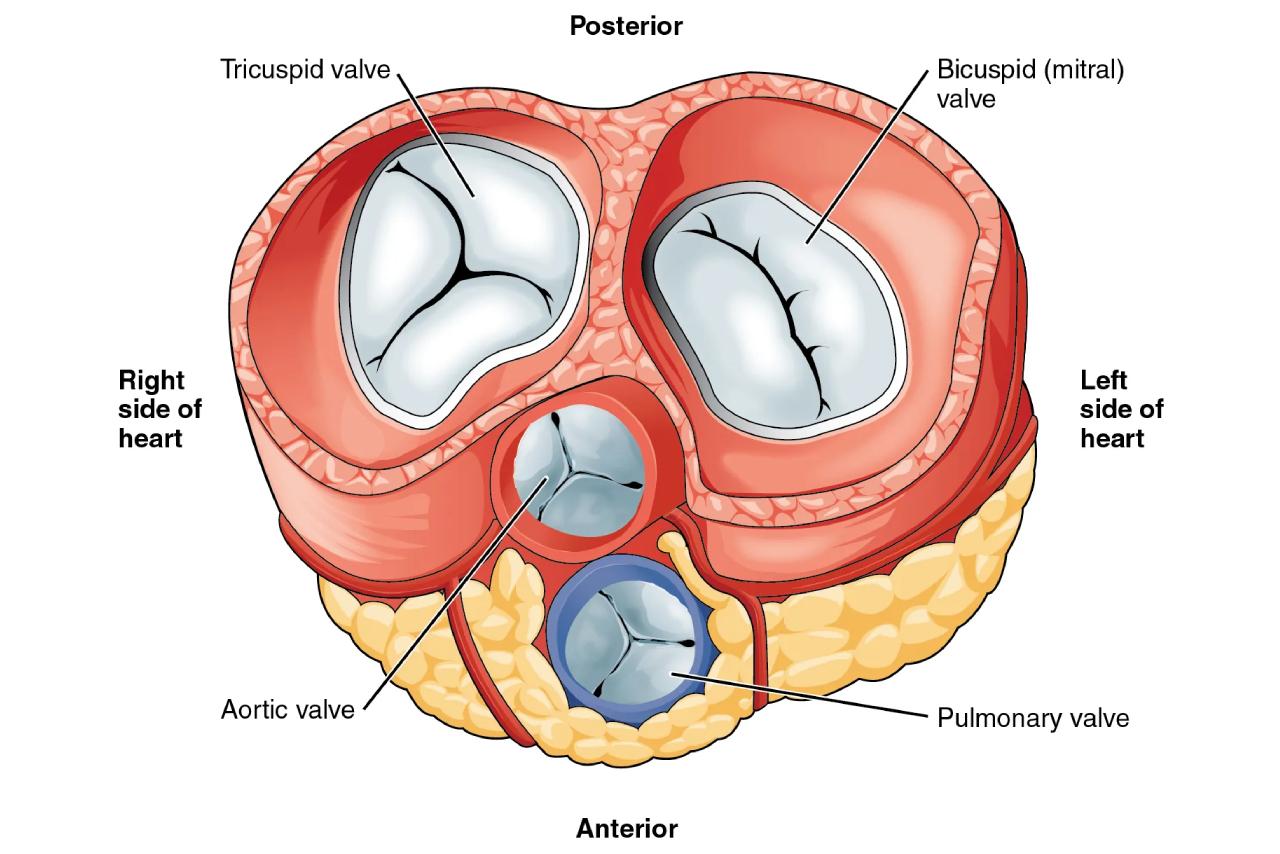
Valves located between the chambers of the heart keep blood flowing through the heart in only one direction.
There are four heart valves named the:
- Mitral valve
- Tricuspid valve
- Aortic valve
- Pulmonary valve
The valves open when blood is pumped forward out of the heart chambers; then close to prevent blood from flowing backwards.
When a heart valve becomes diseased (not working correctly), it may not open or close as it should, and the heart has to work harder to pump blood out to the rest of the body.
The coronary arteries
The heart muscle has blood vessels on its surface called coronary arteries. These arteries supply blood, oxygen, and nutrients to the heart muscle, which needs oxygen-rich blood to function. Below is a picture of the different coronary arteries that wrap around the surface of the heart.
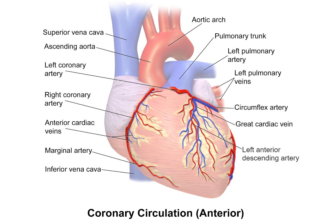
Blausen Medical Communications, Inc., CC BY 3.0 (https://creativecommons.org/licenses/by/3.0), via Wikimedia Commons. The "Anterior interventricular artery" label has been changed to "Left anterior descending artery."
If there is a narrowing or blockage in one of the coronary arteries, the nutrients in the blood will have a hard time reaching the heart muscle. Decreased nutrients and oxygen to the heart muscle will impair the heart's ability to pump blood to the rest of your body.
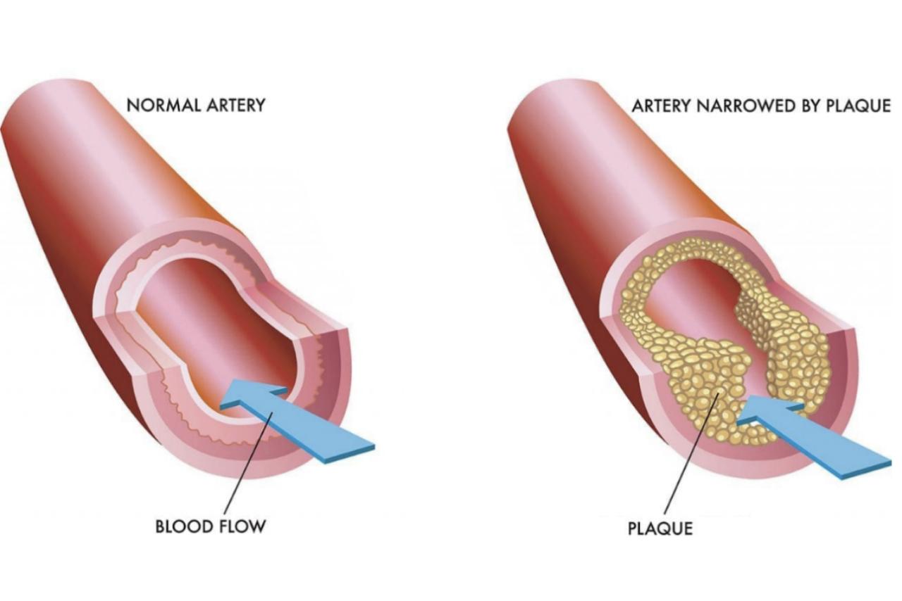
Coronary Artery Disease
Coronary Artery Disease (CAD) is the most common form of heart disease. It happens when one or more coronary arteries become blocked by a buildup of cholesterol fats, often called “plaque.” The vessels become narrow and stiff, affecting the blood supply to the heart.
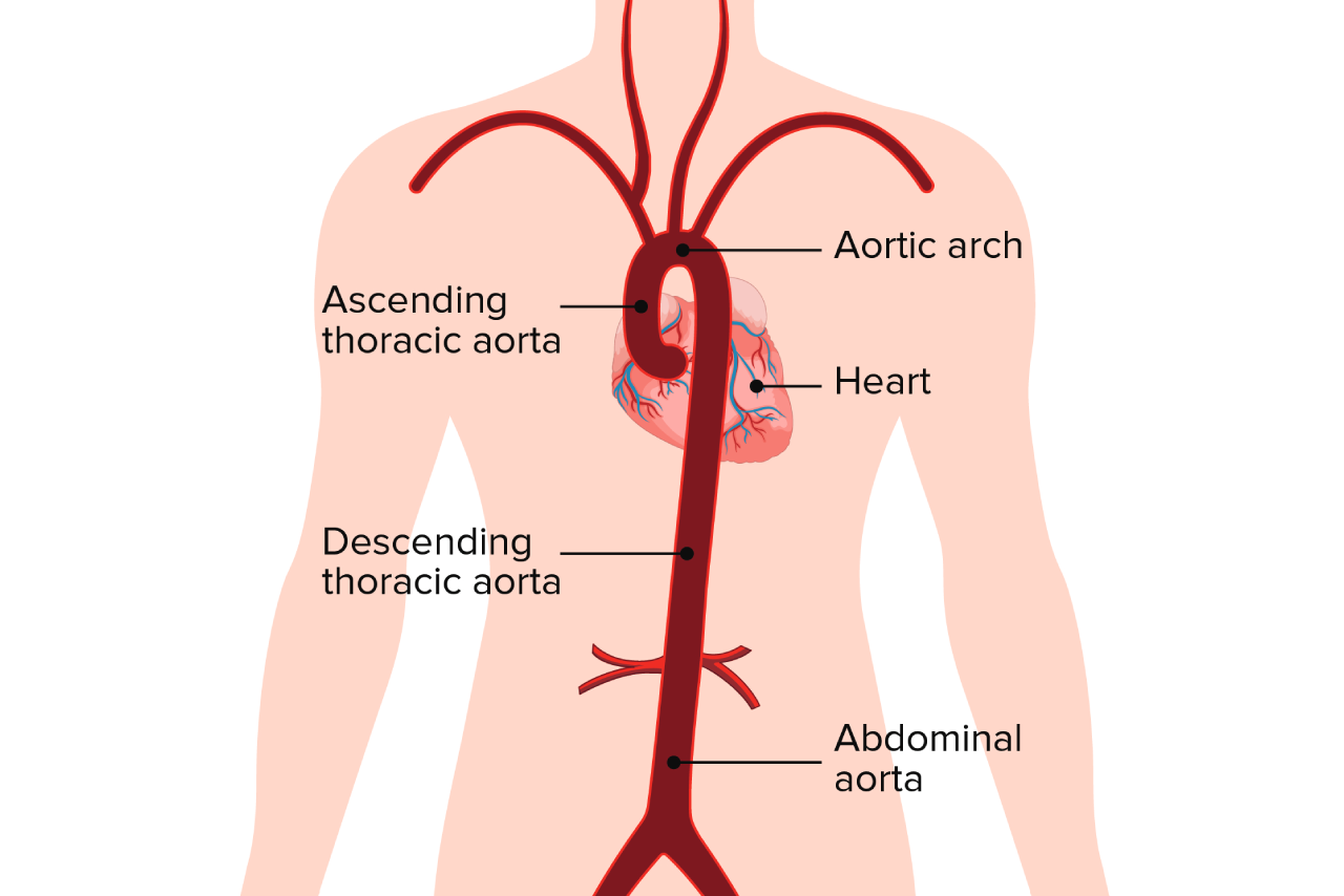
The aorta
The aorta is the largest artery in the body and is the main blood vessel that carries oxygenated blood from the heart to the rest of the body.
Aortic disease
People with aortic disease usually have no symptoms. Aortic disease is often found unexpectedly through imaging tests performed for other reasons (i.e. chest X-rays, CT scans, MRI scans or echocardiograms).
Aortic dissection
An aortic dissection occurs when a tear occurs in the aorta's inner layer. When a tear occurs, blood flows between the layers of the aorta and creates a false lumen (a false channel) for blood to flow. If too much blood flows through the false lumen instead of the true lumen, blood may not be delivered properly to the rest of the body. The area of the tear may also enlarge, and people may develop an aneurysm in addition to the dissection.
If you have any questions about your surgery, or your specific heart condition, please speak with your health-care team.
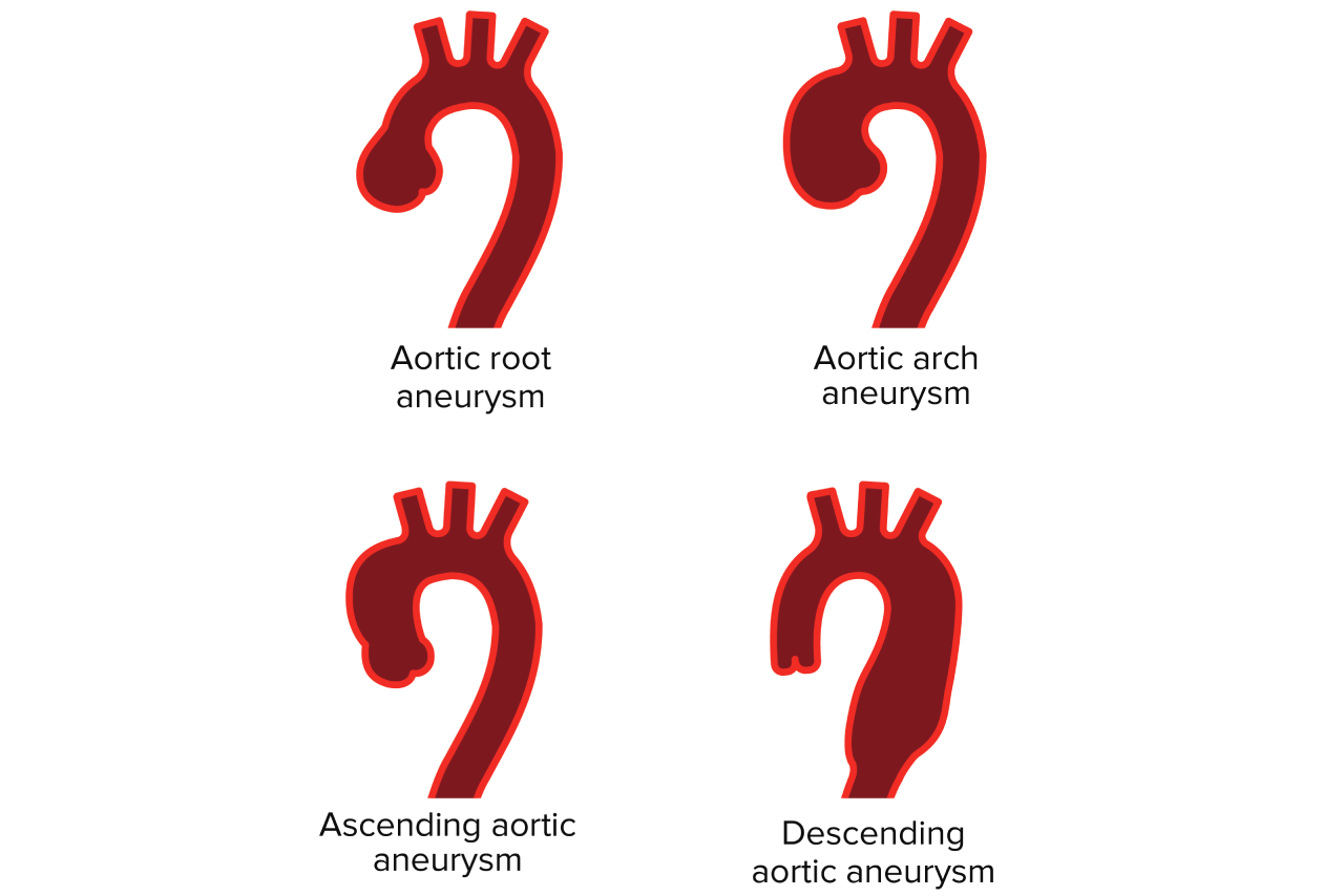
Aortic aneurysm
In aortic disease, the wall of the aorta becomes weak, and a part of the aorta becomes enlarged. The enlarged area is called an aneurysm. If an aneurysm occurs in the part of the aorta running through the chest, it is called a thoracic aortic aneurysm. If it occurs in the part of the aorta running through the abdomen, it is called an abdominal aortic aneurysm.
Depending on the size of the aneurysm, the aorta may be at risk of bursting or tearing. If the aorta bursts or tears, this can cause life-threatening bleeding and affect blood flow to the rest of the body. People with aortic aneurysms require regular follow-up and imaging to monitor the size of the aneurysm. Such follow-up and monitoring are organized through the Thoracic Aortic Disease Program at VGH. If you have a family history of aneurysms or are suspected of having a genetic condition, you may also be referred to a genetic specialist or other specialists. If the size of the aneurysm increases to the point that the risk of bursting becomes concerning, your health-care team will recommend repair.